Label C and the blue circle. By covering your first set of labels you can.

Lab 17 Figure 17 1 Pelvis Diagram Quizlet
Refer urgently to hospital B17.
. Choose your answer to the question and click Continue to see how you did. 4 chamber view that is zoomed in on heart. The anatomy coloring book wynn kapit.
De 2013 Practice for Lab practical 2. First a posterior and anterior view. Call for additional help if possible for mother.
The legs are the lower limbs of the human body that provide support and stability in addition to allowing movement. The acromial region is where the shoulder bones are found. The double lines indicate where bone names are required.
After you have studied the bones in lab label the drawings as a self-test. Diagnostic value and limitations of CT in detecting rib fractures and analysis of missed rib fractures. The names for parts and surface markings of bones are to be written on the single lines ____.
The pubic bones of both pelvis halves are connected via narrow bony skirts that originated at a rather high position on the rear side and continued downwards to a point low on the front side of the shaft. The sacral region is at the end of the spine directly above the buttocks. Perception gives information on the pains.
The lumbar region is the lower back. The head area is the cephalic region. Know cardiac cycle and events terms such as systole and diastole and others related.
On the front edge of the ischial shaft an obturator. Histopathologic study showed a. This is the impression made by the overlying right common iliac artery.
The ischium is S-shaped in side view showing at the transition point between the two curvatures a rough boss on the outer side. Roof-lizard is a genus of herbivorous four-legged armored dinosaur from the Late Jurassic characterized by the distinctive kite-shaped upright plates along their backs and spikes on their tails. Check more flip ebooks related to Bab 6 Sokongan pergerakan dan pertumbuhan of NuRuLhUdA ShAmSuDiN.
It searches only titles inclusions and the index and it works by starting to search as you type and provide you options in a dynamic dropdown list. 4 chamber view which includes view of entire chest. Anatomy of the Heart.
However pain is more than a sensation or the physical awareness of pain. Anatomy And Physiology Bones Lab Test fullexams com. The discomfort signals actual or potential injury to the body.
Moderate-sized pleural effusion on the right. CT of the abdomen and pelvis was performed without oral or IV contrast material per physician request. 74150-26 J90 C.
As the official journal of the Society of Interventional Radiology JVIR is the peer-reviewed journal of choice for interventional radiologists radiologists cardiologists vascular surgeons neurosurgeons and other clinicians who seek current and. M14671 - M16679 M1469. Right ventricular outflow tract showing branching of the PA.
Free Practice Test Instructions. A 2-month-old girl presented with a lesion on her tongue that had evolved over 2 weeks. It also includes perception the subjective interpretation of the discomfort.
Middle right after 24 hours of thrombolysis a bandlike obstruction is seen. Charcots joint ankle and foot. Anatomy and physiology 1 final exam multiple choice quizletAnatomy is the study of the bodies of people and other animals.
CT OF ABDOMEN AND PELVIS. Simplify your Study of Anatomy Physiology. If transfer not possible allow labour to continue.
Instruct assistant family staff to position the womans buttocks higher than the shoulder. Leg Muscle Anatomy. Full length article Open Access.
You may use this feature by simply typing the keywords that youre looking for and clicking on one of the items that appear in the dropdown list. Stegosaurus ˌ s t ɛ ɡ ə ˈ s ɔːr ə s. Far left after stent placement image shows.
The thoracic region is the upper part of the back and chest. Middle left after 12 hours of catheter-directed thrombolysis an obstruction at the left common iliac vein is evident. Full length article.
No previous CT scans for comparison. Interested in flipbooks about Bab 6 Sokongan pergerakan dan pertumbuhan. Clinical impact of allergy and pre-medication in CT studies with.
Maximise the size of the pelvis avoid positions that restrict the movement of pelvic bones eg. AnatomyZone is the leading resource for simple and concise 3D anatomy tutorials with over 200 videos and a new range of interactive 3D anatomy models. Label the items shown in the posterior view of the male reproductive structures.
Push the head or presenting part out of the pelvis and hold it above the brimpelvis with your hand on the abdomen until caesarean section is performed. Resting space between contractions also allows the baby to. The osteology of the lower limb is particularly detailed with 3D view and patterns of bone structures and muscle insertions and ligaments of the hip bone the femur the patella tibia the fibula tibial plateau the tibial pilon the foot talus calcaneus cuboid cuneiform bones metatarsal bones phalanges proximal middle and distal.
Match the technique of birth control to. The interstitial cells of the testes produce. A study based on early CT and follow-up CT as the reference standard.
Surgical resection of the lesion was performed. Secondary malignant neoplasm of bone and bone marrow. Discover 15 secret strategies that will raise your score on any multiple choice exam regardless of the subject.
Female Pelvis With Upper Thighs Firm Fundus Boggy Fundus Simulated Blood Peri Pads 5 Baby Powder 1 Femur Fibula Patella and Tibia Bones Lateral and Medial Meniscus Quadriceps Femoris Tendon Anterior Cruciate Fibular and Tibial Collateral Patellar and Posterior Meniscofemoral Ligaments 1. Pain Definition Pain is an unpleasant feeling that is conveyed to the brain by sensory neurons. The physical examination revealed a tumor hard in consistency located in the posterior midline of the tongue with a base of approximately 1 cm and 2 cm in length.
Dont sit on the back of the pelvis Maximise the ability of the baby to rotate A mobile mother offers lots of opportunities for baby to move water immersion is good for this. Far left view of the entire pelvis demonstrates iliac occlusion. Label the structures seen in the midsagittal section of the female pelvis.
Fossils of this dinosaur have been found in the western United States and in Portugal where they are found in Kimmeridgian- to early. The different joints of the carpus including the distal radioulnar joint the radiocarpal midcarpal and carpometacarpal articulations. Malignant neoplasm of scapula and long bones of upper limb short bones of upper limb pelvic bones sacrum and coccyx long bones of lower limb or short bones of lower limb.
The legs include the upper leg knee lower leg ankle and. Do not spend your precious lab time labeling these drawings this is work you can do at home. Lt ventricular outflow tract 5 chamber view which shows the relationship of the Aorta to the ventricular septum.
Share Bab 6 Sokongan pergerakan. View flipping ebook version of Bab 6 Sokongan pergerakan dan pertumbuhan published by NuRuLhUdA ShAmSuDiN on 2020-07-15. Daniel McNabney and DeLoris Hesse.
M8430x - M8438x Stress fractures. Cine clip of the 4 chamber view sweeping through the outflow tracks. JVIR published continuously since 1990 is an international monthly peer-reviewed interventional radiology journal.
Tamoxifen and raloxifene act by. Label the structures seen in the midsagittal section of the female pelvis. The anatomy of the bones of the wrist subdivided into radius ulna carpal bones scaphoid lunate pisiform triquetrum hamatum capitatum trapezoid and trapezium and the metacarpal bones.
The posterior view shows the following regions. Quick search helps you quickly navigate to a particular category.
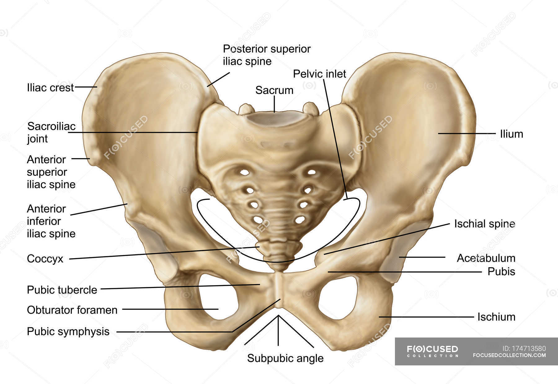
Anatomy Of Human Pelvic Bone With Labels Osteology Biology Stock Photo 174713580

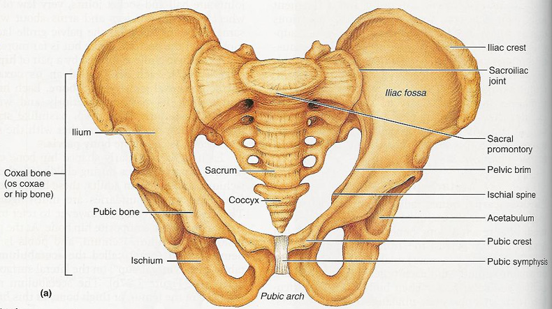
The Pelvic Girdle And Pelvis Anatomy And Physiology I

Human Skeleton System Pelvis With Labels Anatomy Stock Photo Download Image Now Istock
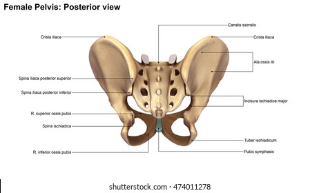
Skeleton Pelvis Posterior View 3d Illustration Stock Illustration 474011278
Pelvis Anatomy Recon Orthobullets
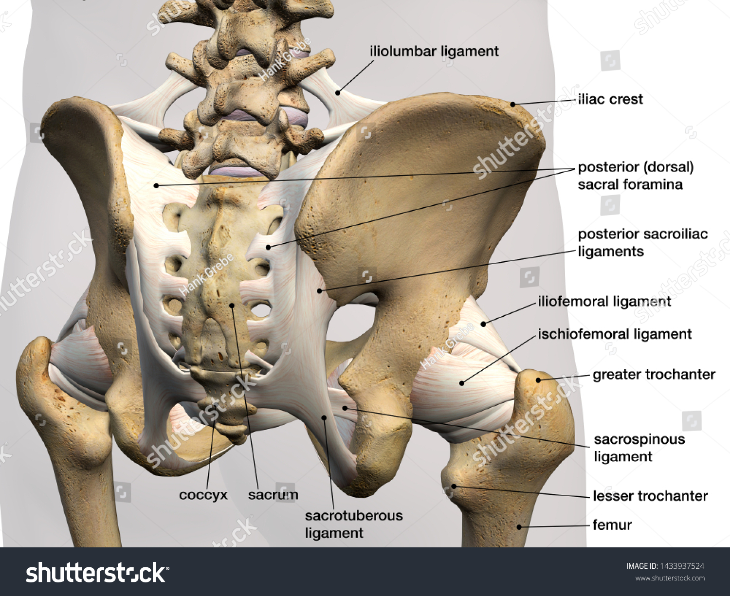
Pelvic Hip Bones Ligaments Labeled Posterior Stock Illustration 1433937524

Anatomy 2017 Unit 3 Label The Bones Of The Pelvic Girdle Anterior View Diagram Quizlet

Coxal Pelvic Bone Posterior View With Labels Appendicular Skeleton Visual Atlas Page 18 Anatomy Flashcards Medical Anatomy Pelvic Bone
0 comments
Post a Comment